Optical Microscopy and Wavefront Shaping
Group leader: Alexander Jesacher
Our group develops new methods for deep-tissue optical microscopy and complex light shaping, combining adaptive optics and computer-generated holography to extend the limits of what can be visualized inside biological specimens.
Deep-Tissue Imaging via Nonlinear Microscopy and Adaptive Optics
Optical microscopy allows us to visualize cells and tissues with sub-micrometer resolution and rich functional contrast. In our group, we develop advanced imaging methods that extend the current limits of optical microscopy, with the goal of increasing imaging depth, improving image quality inside scattering tissue, and extracting more information from biological samples.
A central component of our work is dynamic wavefront shaping, which enables precise control of light fields at megapixel resolution and kilohertz update rates. These technologies allow us to correct distortions introduced by biological tissue and to restore diffraction-limited imaging performance deep inside complex specimens.
Multi-Photon Microscopy
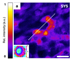
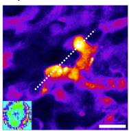
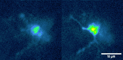
Understanding how cells function inside intact tissue requires imaging techniques that remain effective even in strongly scattering environments. Multi-photon microscopy meets this challenge by using ultrashort laser pulses in the near-infrared spectral range (typically 800–1300 nm), where light penetrates deeper into biological tissue.
Because nonlinear optical signals are generated only at the focal point, multi-photon microscopy naturally provides optical sectioning and enables high-contrast three-dimensional imaging hundreds of micrometers deep—and in favorable cases approaching millimeter depths—in challenging specimens such as brain tissue or organoids.
Adaptive Optics
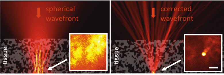
Imaging at even greater depths requires compensating optical distortions introduced by tissue inhomogeneities. In our research, we address this challenge using adaptive optics and dynamic wavefront shaping.
By shaping the excitation wavefront before it enters the specimen, aberrations and scattering effects can be partially compensated, allowing a tight focus to be restored deep inside the tissue. This improves signal strength, resolution, and contrast in otherwise inaccessible regions.
Complex Light Shaping and Computer-Generated Holography
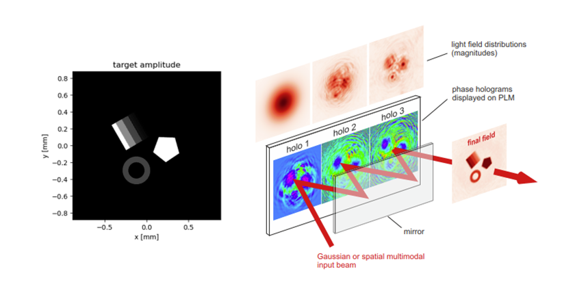
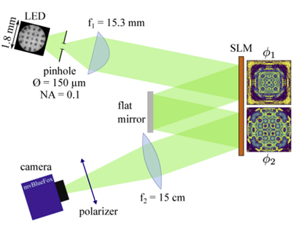
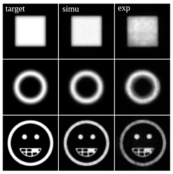
Computer-generated holography (CGH) allows us to design complex light fields numerically and generate them experimentally using programmable diffractive optical elements. This enables precise control over the amplitude and phase of light and opens new possibilities for adaptive optics, structured illumination, and point-spread-function engineering in advanced microscopy.
Selected publications
Nam K., Borozdova M., Park J., Jesacher A., Fast converging iterative wavefront sensing for scatter compensation in multi-photon fluorescence microscopy, J. Phys. Photonics, DOI:10.1088/2515-7647/adeb1f
Maloberti J. G. et al., Joint estimation of point spread function and molecule positions in SMLM informed from multiple planes, Biomed. Opt. Express, 1310, DOI:10.1364/BOE.551278
Muñoz-Bolaños J. et al., Confocal Raman Microscopy with Adaptive Optics, ACS Photonics, DOI:10.1021/acsphotonics.4c01432
Sohmen M., Borozdova M., Ritsch-Marte M., Jesacher A., Complex-valued scatter compensation in nonlinear microscopy, Phys. Rev. Applied, DOI:10.1103/physrevapplied.22.044036
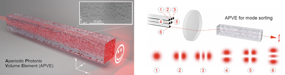
Barré N. et al., Direct laser-written aperiodic photonic volume elements for complex light shaping with high efficiency: inverse design and fabrication, Advanced Photonics Nexus, 036006-036006, DOI:10.1117/1.APN.2.3.036006
May M. et al., Fast holographic scattering compensation for deep tissue biological imaging, Nature Communications, 4340, DOI:10.1038/s41467-021-24666-9