Acoustically-mediated Optical Tomography
Leaders: Monika Ritsch-Marte, Gregor Thalhammer-Thurner
We have pioneered acoustic micromanipulation to enable optical tomography of biological samples such as organoids and cancer spheroids. These specimens are often too optically opaque for single-sided illumination, making conventional 3D imaging challenging or inaccessible. While holographic optical tweezers can rotate individual cells for tomographic studies, extending this approach to larger samples would require intensities that risk overheating. Acoustic radiation forces offer a preferred alternative, enabling non-contact levitation and controlled reorientation for multi-view imaging.
- Kvåle Løvmo M., Moser S., Ritsch-Marte M. et al., Ultrasound-induced reorientation for multi-angle optical coherence tomography, Nature Communications 15, 2391, DOI:10.1038/s41467-024-46506-2
- Moser S., Ritsch-Marte M. et al., Optical tomography reconstructing 3D motion and structure of multiple-scattering samples under rotational actuation, Optica 12, 594-603, DOI:10.1364/OPTICA.550450
Ultrasound fields facilitate the levitation of millimeter-sized objects, including zebra fish larvae, enabling contactless manipulation. With an arrangement of several transducers from different directions, we have realized a setup that allows us to reorient such large specimens in a controlled manner and thus allows us to easily obtain images from different views.
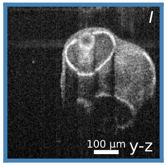
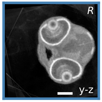
Ultrasound-induced optical coherence tomography
By combining several optical coherence tomography (OCT) images taken from diverse perspectives, we drastically improve the quality of the resulting 3D reconstruction, exemplified here of by zebra fish larva. Compared to a single OCT scan, the ULTIMA-OCT reconstruction provides artifact-free access to the entirety of the specimen with exceptional detail.
Optical tomography is a method to reconstruct the 3D structure of unstained specimens. For samples with strong scattering, such as cell clusters, a single image is not sufficient, but many images from largely different view or illumination angles are necessary. This typically requires a cumbersome mounting of the sample on a rotatable holder. To overcome this hurdle we devised a non-invasive method to hold and rotate specimens using ultrasound waves, which allows for a gentle, contactless manipulation of living specimens. However, with such a device the rotation axis and rotation speed bears some uncertainties since they depend on the complex particle shape. Due to these uncertainties conventional reconstruction methods cannot be applied as they rely on precise knowledge about the particle position and orientation.
In view of this challenges we developed a method to also reconstruct the motion of the sample together with its structure from a series of holograms. We demonstrated that this method is able to deliver accurate, high resolution structural information of cells and cell clusters.